The Latest Innovations in Dental Imaging Technology Transforming Modern Dentistry
Curious about how these tools affect your dental care? Imaging advancements like cross-polarisation OCT and advanced scanning mean earlier detection of decay and disease, which helps you avoid bigger dental problems down the line. If you’ve ever worried about sitting through a messy mould for impressions, digital 3D scanning now removes the need for uncomfortable trays in your mouth.
You can use these technologies to get the most out of your next dental check-up. Ask your dentist if they use CBCT or digital scanners, and see how these innovations make diagnoses and treatment planning more precise. New imaging tools can even help visualise your smile’s potential before any procedure begins, making your dental care more predictable and personalised than ever.
Overview of Modern Dental Imaging Technology
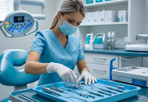
Modern dental imaging equips you with fast, detailed insights into oral health. Today’s advances boost diagnostic accuracy, patient comfort, and efficiency across a range of dental procedures.
Digital Radiography
Digital radiography replaces traditional films with electronic sensors, letting you view images instantly. You can zoom in, enhance contrast, and share results within seconds, which speeds up diagnosis and treatment planning. This technology emits less radiation than film X-rays—often up to 80% lower—making it a safer choice for routine dental care.
You’ll find three main types in use: direct digital (using sensors), indirect digital (using plates), and semi-direct methods. Dentists can store and access images digitally, simplifying long-term record keeping and sharing with other providers. Routine bitewings, panoramic, and periapical views all benefit from digital clarity and efficiency.
Practical tip: Always request digital copies of your X-rays. This way, you can carry your dental history if you change providers or need a specialist consult.
3D Imaging Advancements
3D imaging, especially Cone Beam Computed Tomography (CBCT), has transformed complex dental diagnostics. With CBCT, you get a precise three-dimensional view of teeth, jawbone, nerves, and soft tissue, all from a single scan lasting under a minute.
This technology is crucial for planning dental implants, evaluating jaw disorders, and detecting hidden problems that two-dimensional images can miss. For example, CBCT scans help dentists avoid nerves or sinuses during implant procedures, reducing complications. The approachable size of CBCT machines also means you can often have scans completed during a standard dental appointment.
Did you know? CBCT machines typically use a lower radiation dose than conventional medical CT scans, making them well suited for dental imaging. Always inform your dentist if you are pregnant, as a precaution, before undergoing any 3D scan.
Breakthroughs in 3D Dental Imaging
Recent advancements in 3D dental imaging offer clearer visualisation, improved diagnostic ability, and greater precision in creating dental restorations. Modern scanners and imaging devices have shortened chair times and often reduce the need for traditional impressions.
Cone Beam Computed Tomography
Cone Beam Computed Tomography (CBCT) delivers 3D images of teeth, bone, soft tissues, and nerve pathways in a single scan. You benefit from fast scanning times, usually under a minute, and detailed images at various resolutions.
CBCT is particularly effective in complex cases like implant planning, endodontics, and jaw joint analysis. You avoid unnecessary exposure to radiation, as dental CBCT units typically use lower doses than hospital CT scanners. Most machines feature small footprints and operate in regular dental surgeries.
Key Features of CBCT:
- 3D imaging for accurate treatment planning
- Rapid acquisition times (as quick as 20 seconds)
- High detail with voxel sizes as small as 0.08 mm
Tip: Ask your dentist if a focused, limited-scope CBCT scan can answer your clinical needs. This approach often lowers both time and cost.
3D Intraoral Scanners
With 3D intraoral scanners, you no longer need traditional impression materials that can cause discomfort or inaccuracies. Scanners use advanced optics to create a digital replica of your teeth and bite, which is visible instantly on screen.
You benefit from quicker procedures—many full-arch scans complete within two minutes. Digital files streamline clear aligner and crown creation, enhancing the fit and reducing the number of adjustments.
Applications of 3D Intraoral Scanners:
- Digital models for orthodontic aligners, crowns, and bridges
- Direct communication with dental laboratories via secure digital transfer
- Monitoring wear on teeth or changes in dental health over time
Fun Fact: Some scanners can even match tooth shade automatically, helping improve restoration aesthetics with less guesswork.
Artificial Intelligence in Dental Imaging
Artificial intelligence (AI) is driving rapid changes in dental imaging. You benefit from sharper diagnostics, streamlined workflows, and more reliable results every time you visit your dentist.
AI-Powered Diagnostics
AI algorithms analyse dental images with impressive precision, often identifying details that even experienced clinicians might miss. Deep learning models, trained on thousands of annotated images, can recognise early signs of tooth decay, periodontal disease, and bone loss. This means you may receive earlier interventions, leading to less invasive treatments and better long-term oral health.
Dentists use AI to compare your scans over time, flagging changes automatically. Some systems can detect hidden caries between teeth, which are difficult to spot with the naked eye. These advancements are not just limited to X-rays; AI now supports interpretation of 3D cone beam computed tomography (CBCT) and panoramic images as well.
Tip: Always ask your dental practitioner if they use AI-supported imaging for the highest diagnostic accuracy.
Automated Image Analysis
AI can now automate the time-consuming process of analysing and segmenting structures in dental images. The software can pinpoint features such as nerve locations, tooth roots, and bone density patterns in just a few seconds. This speeds up case planning, especially for implants or orthodontic work.
Dentists can use the automatic measurements provided by AI software to plan treatments more precisely. For instance, AI-based tools can measure jaw length in inches (and centimetres), identify impacted teeth, or estimate bone width in fractions of an inch (millimetres).
Fun fact: Some practices use AI to generate annotated reports, including diagrams and tables, that you can review on your own device before making decisions about your care.
Enhancements in Imaging Workflow and Patient Care
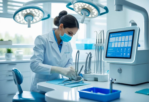
Modern dental imaging technology makes your visits more efficient and comfortable. Managed workflows, better software, and patient-focused designs all reduce appointment time while improving the accuracy of each image.
Improved Imaging Software
Recent upgrades to imaging software streamline diagnosis and treatment planning. Artificial intelligence (AI) now helps spot tooth decay, bone loss, and other abnormalities quickly and accurately. You gain precise results in less time, and dentists can compare past and current scans side by side.
Many systems feature guided workflows that clearly prompt each step, reducing the risk of human error or missed details. For example, with DEXIS™ IS ScanFlow and similar programmes, you can watch as the software processes your scan, ensuring all needed areas are captured.
Customisable reporting lets dentists highlight important findings with notes or diagrams. Integration with practice management systems means your records stay organised and instantly accessible. If you need a referral, your images are easy to share or export, which helps dental teams work together efficiently.
Patient Comfort Innovations
Today’s digital imaging tools focus on making your experience more pleasant. Intraoral scanners no longer need messy, uncomfortable impressions. The handheld devices fit easily in your mouth and complete a full scan in just a few minutes.
Many imaging systems now reduce exposure by using low-dose radiation, which adds a layer of safety. With faster scans and fewer retakes, you spend less time sitting still in the chair. If you find traditional X-rays difficult, digital panoramic units offer an open design, helping minimise any feelings of claustrophobia.
You can also see your scans on a screen in real time, making it easier to understand your treatment plan. Communication improves, and you get a clearer view of what’s happening with your dental health. These advancements make visits much smoother, particularly for children or those with dental anxiety.
Future Directions and Emerging Trends
Dental imaging technology is poised for major transformation, driven by advances in integration and personalisation. These innovations focus on streamlining your dental experience and enhancing diagnostic capabilities for more predictable results.
Integration with Other Digital Dental Technologies
Modern dental imaging is now compatible with a suite of digital dental tools. This integration allows you to benefit from workflows that combine 3D imaging with CAD/CAM systems, digital impressions, and even AI-based diagnostic platforms.
For example, a digital scan of your mouth can now be used in tandem with a cone-beam CT image. This pairing enables precise planning for implants or orthodontic treatment in just one office visit. With cloud-based record sharing, images are instantly accessible across clinics and devices.
Benefits of Integration:
- Faster turnaround for crowns and aligners
- Real-time visual communication between you and your dentist
- Fewer errors due to digital precision
- Simplified appointment scheduling by consolidating digital records
Fun fact: Some practices can now create same-day crowns with digital impressions, cutting lab wait times from weeks to hours.
Potential for Personalised Treatment Planning
Personalised dentistry is progressing quickly thanks to cutting-edge imaging. With detailed scans and digital analysis, your dentist can tailor treatment to your exact anatomy, needs, and health history.
AI-powered image analysis can predict risks such as developing cavities or gum problems before symptoms occur. By overlaying digital 3D models from different devices, your dentist can simulate outcomes for treatments from braces to bone grafts.
Personalisation in Practice:
- Adjustable treatment plans that change as your mouth changes
- Simulations help you visualise treatment outcomes
- Fewer unnecessary X-rays due to targeted, need-based imaging
- Higher accuracy in planning surgical guides for implants
Tip: Ask if your dentist uses digital imaging software that allows you to view and discuss treatment options on-screen. This empowers you to make informed decisions and understand every step.
Frequently Asked Questions
Artificial intelligence, digital sensors, 3D printing, improved cone beam CT scans, portable devices, and augmented reality are rapidly transforming dental imaging. You can now access faster, more precise, and less invasive diagnostic tools in dental care than ever before.
How are AI algorithms enhancing dental diagnostic accuracy?
AI software is now analysing X-rays and scans to detect hidden cavities, bone loss, and even early signs of oral cancer. You can benefit from more consistent diagnostic results because the technology highlights suspicious areas that may be missed by the human eye.
Some dental practices are using AI to predict tooth decay risk, track changes over time, and compare your scans against vast image databases. This speeds up second opinions and reduces the likelihood of diagnostic errors.
What are the latest developments in digital intraoral sensors?
Modern intraoral sensors have become slimmer, more comfortable, and much faster. You’ll notice less waiting time for your dentist to view high-resolution images on a computer screen.
Many new sensors deliver images instantly and automatically adjust exposure levels to lower the radiation dose. Some models are even designed to better fit different mouth sizes, including for children.
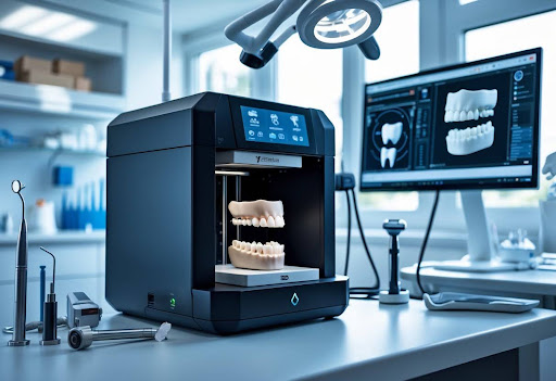
In what ways have 3D printing technologies impacted dental imaging?
3D printing now lets dental teams produce custom surgical guides, orthodontic models, and accurate replicas of jaw structures directly from your digital scans. This means you can have crowns, bridges, or aligners tailored for a precise fit.
Dentists use printable models for treatment planning and patient education. It’s also possible to print models within a few hours, which can save several days compared to traditional methods.
Can you describe the improvements in dental CBCT imaging?
Dental cone beam CT (CBCT) systems now generate clearer, three-dimensional images with less scatter and distortion. You’re exposed to less radiation because imaging protocols are more efficient.
Higher resolution means your dentist can see small cracks, root canals, or sinus pathways in much finer detail. CBCT is now accessible in smaller practices due to reduced size and cost of equipment.
What innovations have been made in portable dental imaging systems?
You can now find handheld X-ray devices that operate on battery power and produce instant images. These systems allow for mobile dentistry in care homes, community clinics, or even remote regions.
Portable imaging is handy for quick checks during emergencies or surgeries. Some devices are just a few pounds (around 2 to 3 pounds, or 900g to 1.4kg), making them easy to carry from room to room.
How is augmented reality being integrated into dental imaging practices?
Augmented reality (AR) overlays scans, X-rays, or 3D models directly onto your view of a patient’s mouth during examination or treatment. Dentists can see virtual guides or treatment plans in real-time to improve precision.
A few AR platforms now offer interactive education where you can visualise planned procedures superimposed onto your own teeth. This helps with understanding what to expect during your dental treatment.